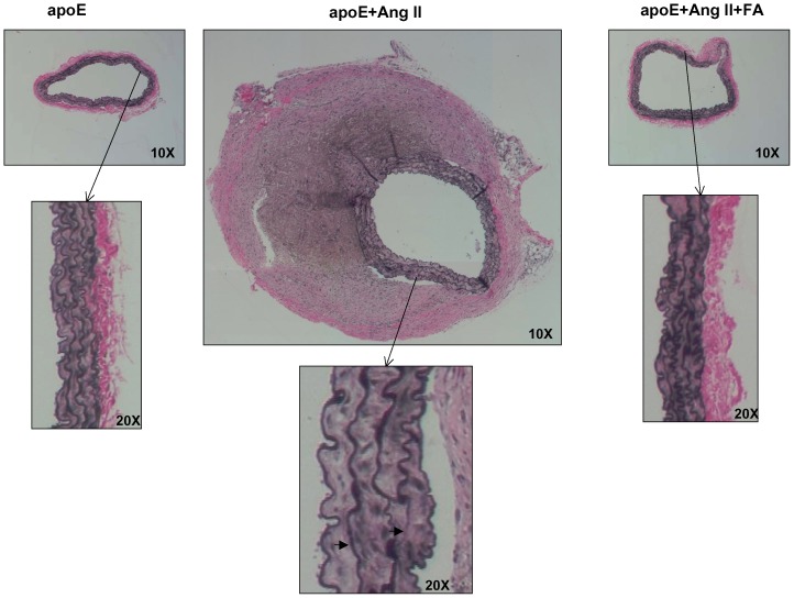
Figure 4. Folic acid reduces matrix degradation in Ang II-infused apoE null mice.
Abdominal aortas were collected from sham operated (left column), Ang II-infused (center column), and Ang II-infused and folic acid (FA, right column) treated apoE null mice 4 weeks after infusion. Tissues were then sectioned and stained for VVG; the right arrow in the VVG sections points to a breakdown of elastin fiber, while the left arrow points to a flattening of elastin fiber.

