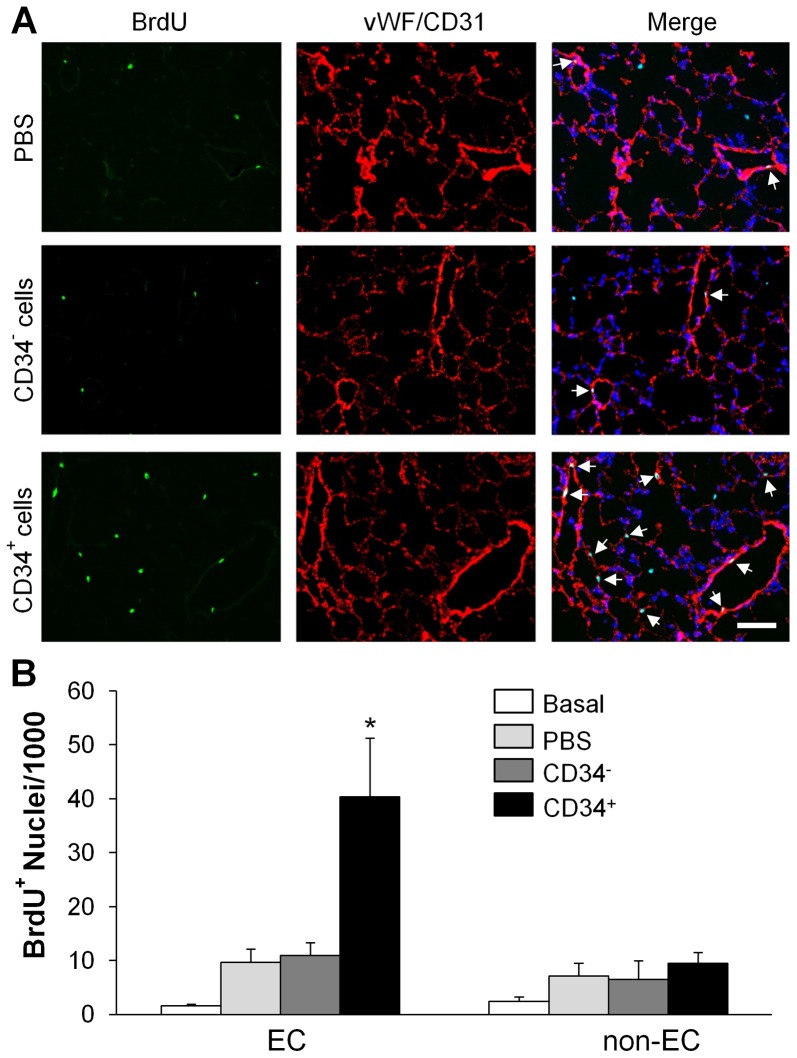
Figure 6. fCB-CD34+ cell treatment-induced endothelial cell proliferation in lungs.
(A) Representative micrographs of immunofluorescent staining. Lung tissues were collected at 52 h post-LPS challenge, sectioned and immunostained with anti-BrdU (green) and anti-vWF/CD31 (red) antibodies. Nuclei were counterstained with DAPI (blue). Arrows indicate proliferating EC. Scale bar: 50 µm. (B) Quantification of BrdU-positive EC (vWF+ and/or CD31002B) and non-EC (vWF− and/or CD31−). Data are expressed as mean ± SD (n = 4/group). *, P<0.001 versus PBS or CD34−.

