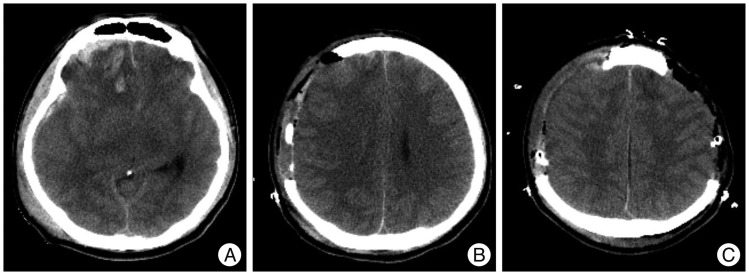
Fig. 2.
Pre- and postoperative computed tomography (CT) scans of an illustrative case. A : Preoperative CT scan showing diffuse brain swelling and obliterated basal cisterns. B : Initial postoperative CT scan showing early decompressive craniectomy (DC). C : Postoperative CT scan showing consecutive bilateral DC at an interval of eight hours.

