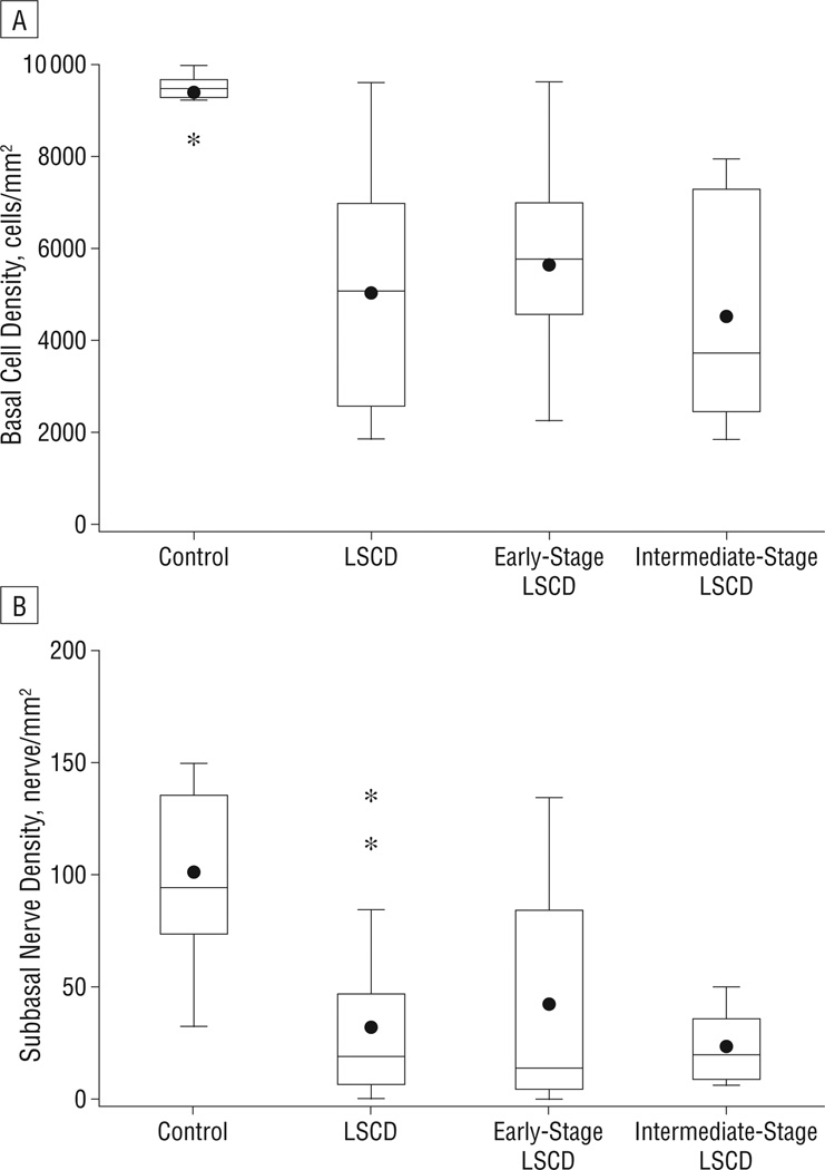
Figure 3.
Box and whisker plot of central corneal basal epithelial cell density (A) and subbasal nerve density (B). There was a significant decrease in basal cell density and subbasal nerve density in all patients with limbal stem cell deficiency (LSCD) and in the subgroups of early-stage and intermediate-stage LSCD compared with controls. *P<.001.
The dots indicate the mean.

