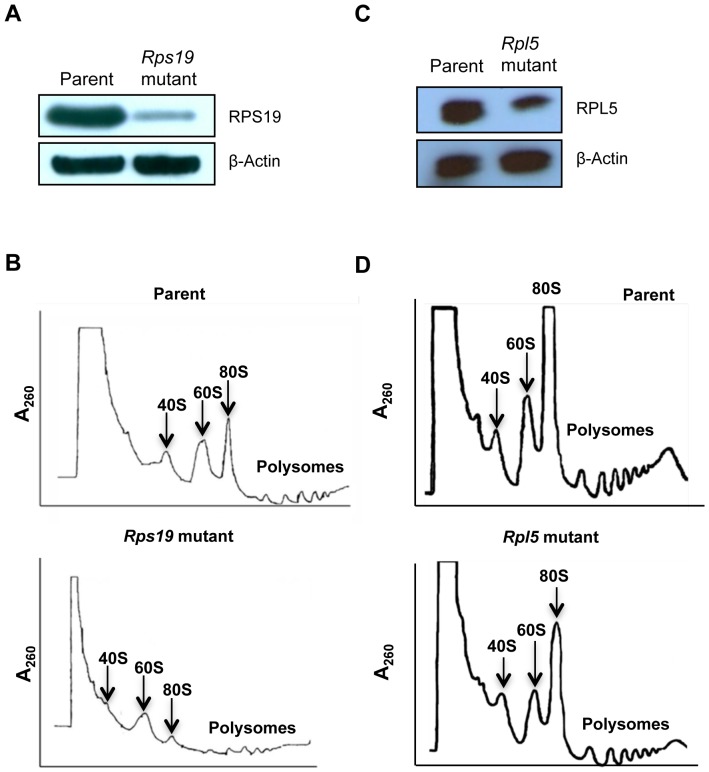
Figure 1. Protein haploinsufficiency and polysome defects in Rps19 and Rpl5 mutant mouse embryonic stem cells.
For immunoblotting, ES cell lysates were separated using gel electrophoresis, transferred to a nitrocellulose membrane, and blotted with antibodies against RPS19 and RPL5. β-Actin was used as a loading control. Rps19 mutant (A) and Rpl5 mutant (C) ES cells showed protein haploinsufficiency (upper panels); β-Actin confirmed similar protein loading for mutant and parent (lower panels). For analyses of polysome profiles, ES cells were incubated in the presence of cycloheximide, lysed, and layered onto sucrose gradients. After ultra-centrifugation, polysome profiles were retrieved using an ISCO density gradient fractionator and UA-6 UV/Vis detector. RPS19 haploinsufficient ES cells (B, lower panel) showed a decreased 40S peak when compared to the parental line (B, top panel). In contrast, RPL5 haploinsufficient cells (D, lower panel) had a decreased 60S subunit compared with the parental cells (D, top panel).

