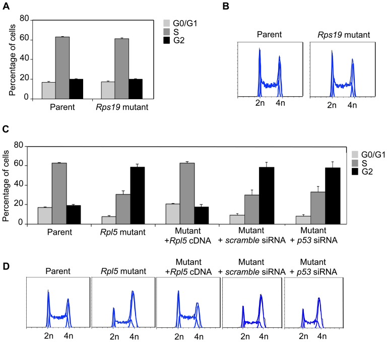
Figure 5. Rpl5 mutant ES cells exhibit a p53-independent cell cycle arrest.
Cell cycle analyses were performed by fixing ES cells with 70% ethanol, followed by staining with PI solution containing RNase A. Quantification of cell cycle phases (A), along with flow cytometry profiles (B) of Rps19 mutant ES cells show no difference, compared to the parent. In contrast, the cell cycle profile of the Rpl5 mutant ES cells exhibited a three-fold increase in the G2 phase with a concomitant decrease in the G1 and S phases, consistent with a delayed G2 phase transition (A, C) (three independent pooled experiments). Stable transfection of the Rpl5 mutant with a vector containing Rpl5 cDNA showed complete correction of the cell cycle defect; however, siRNA knockdown of p53 was unable to rescue the defect (D).

