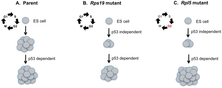
Figure 7. Proposed model suggesting a secondary role for p53 in augmenting erythroid colony formation in mouse ES cell models of Diamond Blackfan anemia.
Wild type mouse embryonic stem (ES) cells can be differentiated into primitive erythroid colonies (A). In the normal setting, colony formation can be further increased by p53 knockdown. (B) Rps19 mutant ES cells exhibit defective primitive erythroid colony formation through an unknown p53-independent mechanism. However colony formation can be augmented by p53 knockdown through a separate p53 dependent pathway. (C) The Rpl5 mutant ES cells show an early cell cycle defect at the ES cell stage that is p53-independent. These cells also exhibit a similar defect in primitive erythroid colony formation through a p53- independent mechanism. p53 knockdown in these cells increases colony formation to a greater degree than the Rps19 mutant cells, for unknown reasons.

