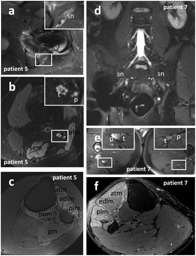
Figure 4. Localization and fascicular distribution of T2-nerve lesion, and denervation pattern of target muscles.
siMRN in patient 5 (a, b, c: transversally orientated T2-weighted TSE with fat-suppression) and patient 7 (d: coronally orientated T2 STIR; e, f: transversally orientated T2-weighted TSE with fat-suppression). Depending on the extent of implant-related artifacts, evaluation of the sciatic nerve may be impaired or even impossible for a certain region (d). Regular depiction of the nerve roots and lumbar pexus in patient 7 (d), but the subtrochanteric sciatic nerve exhibits a peroneally accentuated T2-lesion (e), and the peroneally innervated muscles of the proximal lower leg show signs of denervation (f). In patient 5 the proximal sciatic nerve showed a T2-lesion in proximity of the ischial tuberosity (a), affecting all fascicles of the nerve at level of the hip (a) and thigh (b). Furthermore the denervation of the biceps femoris and semimembranosus muscle of the thigh (b), and the anterior tibialis, extensor digitorum, peroneus longus, popliteus and gastrocnemius muscle of the lower leg (c) indicate affection of the tibial and peroneal division of the sciatic nerve. Abbreviations: atm: anterior tibialis muscle; bfm: biceps femoris muscle; edlm: extensor digitorum longus muscle; gm: gastrocnemius muscle; p: peroneal portion of the sciatic nerve or peroneal nerve; plm: peroneus longus muscle; pom: popliteus muscle; sem: semimembranosus muscle; sn: sciatic nerve; t: tibial portion of the sciatic nerve or tibial nerve.

