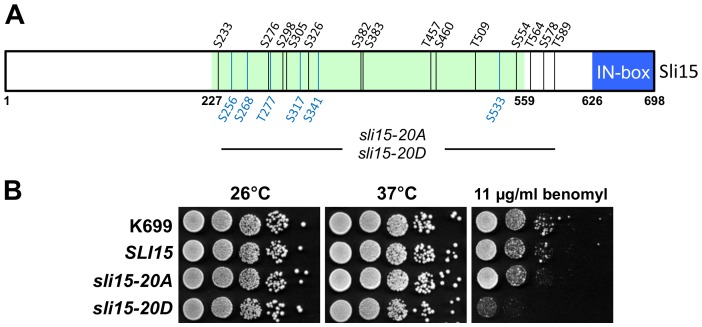
Figure 1. Characterization of the sli15-20A and sli15-20D alleles.
(A) Schematic representation of Sli15 showing the 14 phosphorylation sites mapped in vitro (black) and the six additional sites discussed in the text (blue) that were mutated in sli15-20A and sli15-20D. The microtubule-binding region (residues 227–559) [23] is shaded green and the conserved IN box (residues 626–698) is shaded blue. (B) Equivalent 10-fold dilutions of a wild-type strain (K699) and of sli15Δ::KanMX6 strains with either wild-type SLI15 (VMY30), sli15-20A (VMY148) or sli15-20D (VMY187) integrated at the his3 locus were spotted onto YPAD agar in the presence of absence of benomyl at 11 µg/ml and grown for two days at 26°C or 37°C as indicated.

