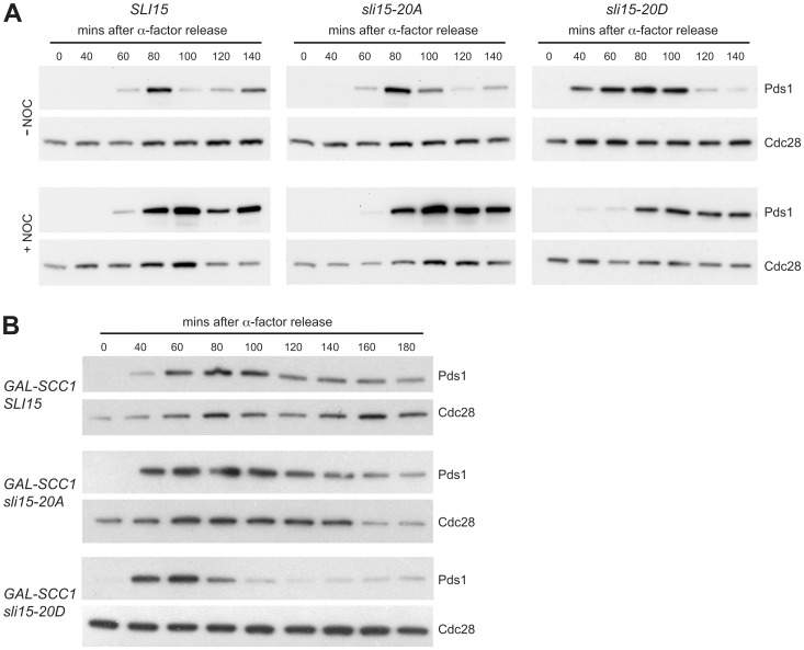
Figure 5. Mimicking constitutive Ipl1-dependent phosphorylation of Sli15 interferes with the checkpoint response to reduced cohesion.
(A) Wild-type SLI15 (VMY194), sli15-20A (VMY162), and sli15-20D (VMY191) strains expressing Pds1-myc 18 were arrested in G1 with α-factor and synchronously released into YPD medium in the presence (+NOC) or absence (−NOC) of 30 µg/ml nocodazole. Samples were collected at the indicated times. Levels of Pds1-myc18 (Pds1) and Cdc28 (loading control) were monitored by immunoblotting using anti-myc and anti-Cdc28 antibodies, respectively. (B) Wild-type pGAL-SCC1 SLI15 (VMY222), pGAL-SCC1 sli15-20A (VMY166) and pGAL-SCC1 sli15-20D (VMY356) cells expressing Pds1-myc 18 were arrested with α-factor for 2 h in medium containing galactose and then released in medium containing glucose to repress pGAL-SCC1. Pds1 and Cdc28 were monitored as described for panel A.

