
Figure 1. No alterations in N-cadherin and β-catenin after knockdown of Dsg2 in melanoma cells.
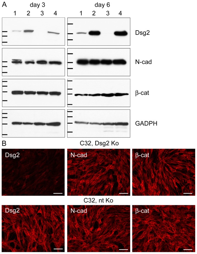
(A) Immunoblots showing efficient Dsg2 depletion in MeWo and C32 melanoma cells. Equal amounts of proteins were loaded. 1: MeWo, Dsg2 siRNA; 2: MeWo, non-targeting (nt) siRNA; 3: C32, Dsg2 siRNA; 4: C32, nt siRNA. In MeWo, Dsg2 reduction was 7.9-fold three days after Dsg2 siRNA transfection and 5.1-fold six days thereafter when the intensity of the bands was normalized against the GADPH immunoblots serving as loading controls. In C32, Dsg2 was 12.7-fold or 122.8-fold reduced. By contrast, protein amounts of N-cadherin (N-cad) and β-catenin (β-cat) were virtually unchanged upon Dsg2 depletion. Molecular weight markers (from top to bottom): Dsg2 immunoblots: 158, 116 and 97.2 kDa (day 3); 212, 158 and 116 kDa (day 6); N-cad immunoblots: 116, 97.2 and 66.4 kDa (day 3); 158, 116 and 97.2 kDa (day 6); β-cat immunoblots: 158, 116, 97.2 and 66.4 kDa (day 3 and 6); GADPH immunoblots: 55.6, 42.7, 34.6 and 27 kDa (day 3 and 6). (B) Immunofluoresence microscopy of Dsg2-depleted (upper panel) and nt siRNA-treated C32 cells (lower panel), showing virtual absence of Dsg2 three days after knockdown. In cells treated with nt siRNA Dsg2 is accumulated at the cell surface and at cell borders. Antibodies to N-cad and β-cat react at cell-cell junctions and along cell borders, in patterns unaffected by Dsg2 contents. Bars: 200 µm.