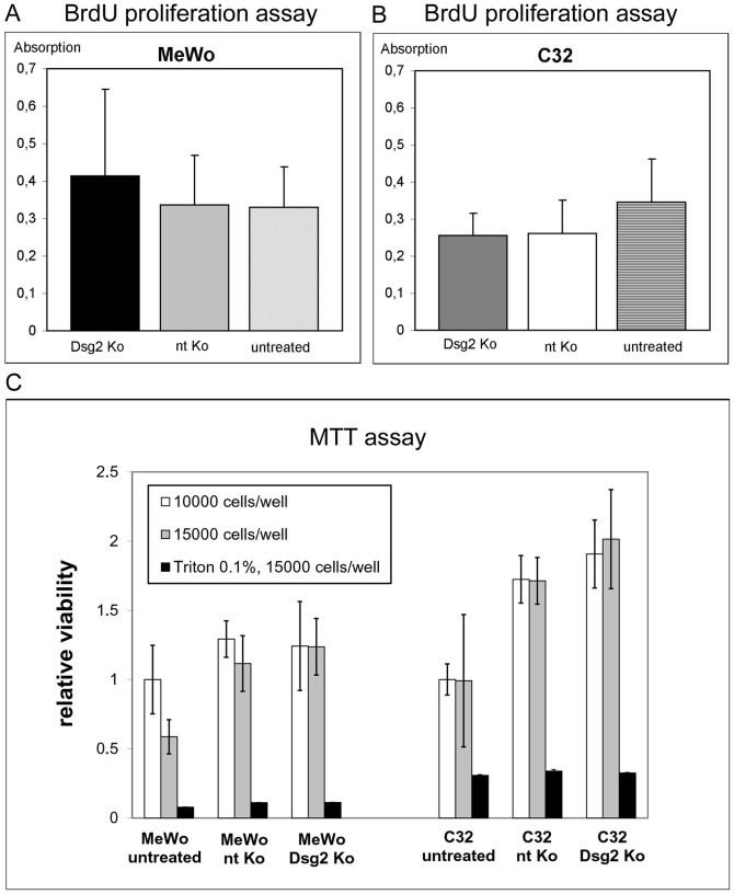
Figure 5. Dsg2 depletion does not significantly influence proliferation and viability of melanoma cells.
(A, B) Bar diagrams showing the absorption rates determined with colorimetric BrdU Cell Proliferation ELISA. Dsg2-depleted MeWo (“Dsg2 Ko”; A) and Dsg2-depleted C32 cells (B) were compared to cells treated with non-targeting siRNA (“nt Ko”) and to untreated cells. Pairwise t-tests gave no significant differences with respect to absorption, indicating similar proliferation rates in all probes. (C) MTT assay comparing the viability of Dsg2-depleted MeWo and C32 cells to non-targeting siRNA-treated and untreated controls at densities of 10000 cells/well (white bars) or 15000 cells/well (grey bars). Quantification and normalization showed no significant differences between Dsg2 Ko and nt Ko cells. Analysis of untreated cells indicated increase of mitochondrial activity induced by the transfection technique. Viability of Dsg2 Ko and nt Ko MeWo and C32 cells is expressed as relative values compared to untreated cells at a density of 10000 cells/well. Treatment with 0.1% Triton X-100 was used as control. Bars: SD.

