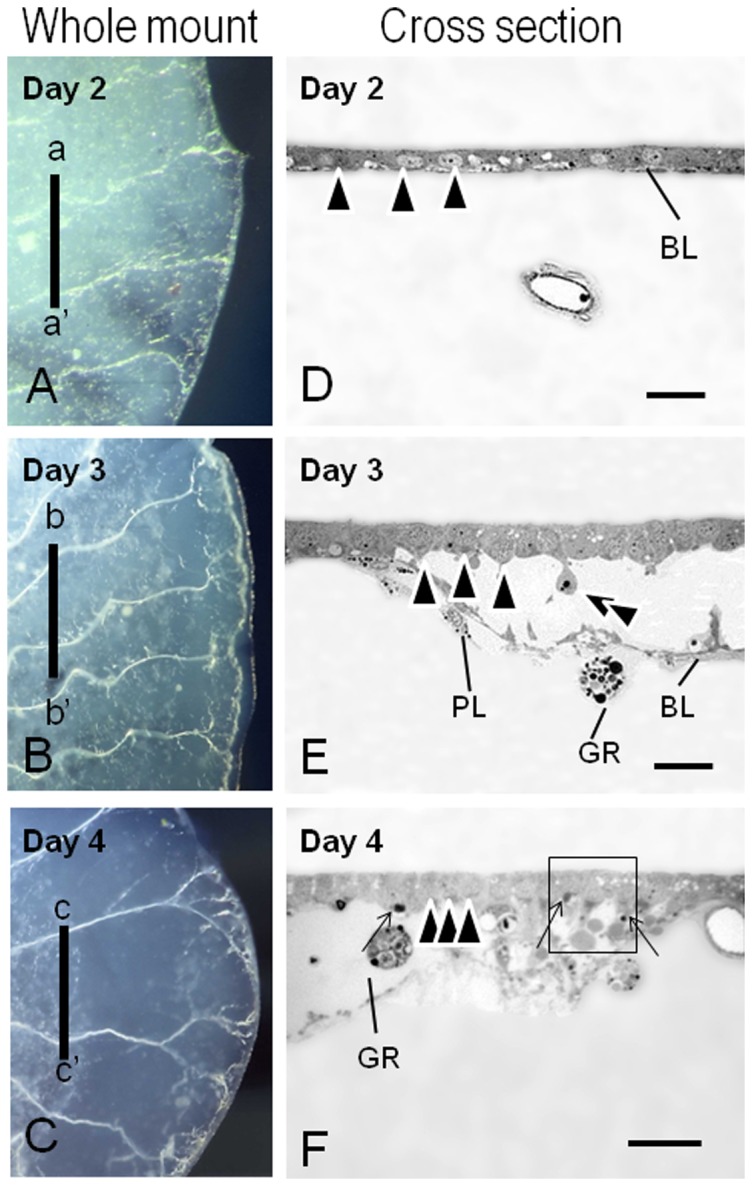
Figure 2. Whole mount images and cross sections of female pupal wings.

Day 2 (A, D), Day 3 (B, E), and Day 4 (C, F) after the beginning of adult development. (A–C) Whole mount of female-pupal wings dissected from a pupa. (D–F) Cross sections of female-pupal wings. a-a′, b-b′, and c-c′ levels of the sections depicted in D–F. The box area in (F) corresponds to Fig. 3(B). An epithelial fragment was degenerating into the wing lumen (B, double arrowheads). An apoptotic body-like structure was also visible (F, black arrows). Note that the position of the epithelial nuclei shrank gradually (D–F, arrowheads). BL, basal lamina; PL, plasmatocytes; GR, granulocytes. Scale bar = 20 µm.
