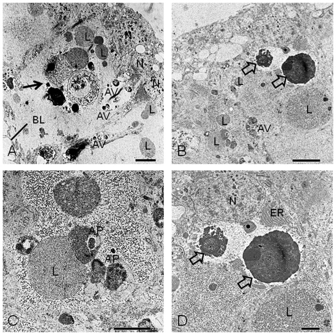
Figure 3. Transmission electron microscope micrographs of the pupal wing epithelium of females.
Cell death of the pupal wing epithelium of females occurred strikingly on Day 4 after the beginning of pupal-adult development. (A) Phagocyte (black arrow) is engulfing fragmented forms of dead cells and lysosomes. (B) Condensed chromatin (open arrows) derived from nuclei are visible. (C) Autophagosome fuses to lysosomes to become an autolysosome. (D) Condensed chromatin and endoplasmic reticulum are visible. N, normal nuclei; ER, endoplasmic reticulum; BL, basal lamina; L, lysosome; AP, autophagosome; AV, autophagic vacuole. Scale bars: 5 µm for A–B and 2 µm for C–D.

