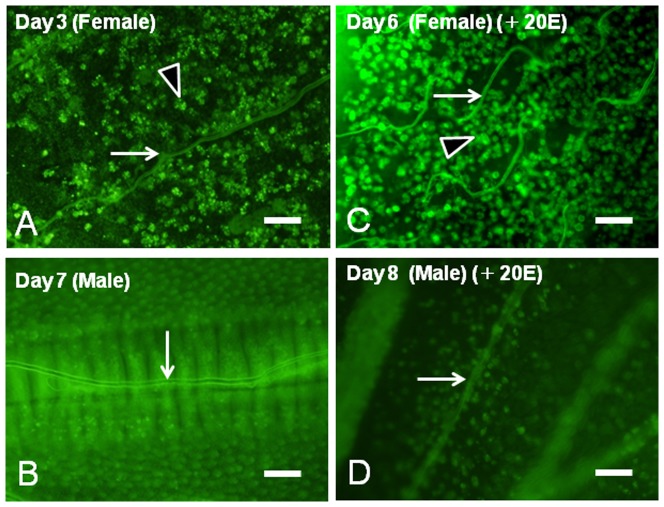
Figure 6. Acridine orange stain detecting the cell death of female pupal wings.
(A) Whole mount of a female pupal wing on Day 4 after the beginning of pupal-adult development. (B) Whole mount of a male pupal wing on Day 7 after the beginning of pupal-adult development. (C) Whole mount of a female pupal wing on Day 6 after injection of 5.4 µg 20E. (C) Whole mount of a male pupal wing on Day 8 after injection of 5.4 µg 20E. Note that the coiling trachea synchronizes with wing degeneration (C, arrow). Most of the large signals in female wings appear to be fragmental nuclei trapped by the phagocytotic actions of hemocytes (A, C, arrowheads). White arrows (A–D) point to trachea. Scale bar = 100 µm.

