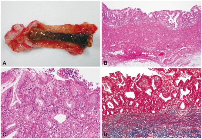
Fig. 3.
Macroscopic and microscopic findings of the stented bile duct (dog 6). (A) Macroscopic examination of the stented bile duct, showing superficial inflammation. (B) Chronic inflammation is confined to the mucosa (H&E stain, ×40). (C) There are a few neutrophils in the lamina propria (H&E stain, ×200). (D) Fibrosis extends to the muscle layer, but smooth muscle bundles are intact (red spindle cells; Masson trichrome stain, ×100).

