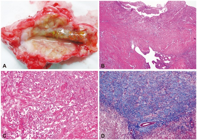
Fig. 4.
Macroscopic and microscopic findings of the stented bile duct with severe inflammatory changes (dog 4). (A) A fully covered self-expandable metal stent became embedded in the bile duct owing to severe epithelial hyperplasia. (B) A low-magnification view reveals transmural inflammation and fibrosis (H&E stain, ×40). (C) Suppurative inflammation with many neutrophils is visible (H&E stain, ×200). (D) Masson trichome stain highlights extensive fibrosis (blue areas; H&E stain, ×100).

