Fig. 5.
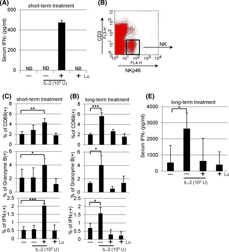
In vivo effects of lunasin. a Lunasin enhances the secretion of IFNγ in the serum. BALB/c mice received single intraperitoneal (IP) injection with PBS (−), IL-2 (1 × 10⁵ U/mouse) without (−) or with (+) lunasin (0.4 mg/kg body weight), or lunasin alone as indicated. Mice were killed 18 h following injection, and blood samples were collected by cardiac puncture. The serum levels of IFNγ were analyzed using ELISA. Data are presented as mean ± SD from 3 mice per group. ND, not detectable. b, c NK activation in vivo following short-term treatment. BALB/c mice received single daily IP injection for 3 consecutive days as indicated. The following day mice were killed, and spleens were collected for analysis of NK activation using flow cytometry. NK cells gated on CD3− NKp46+ populations (b) were analyzed for surface expression of activation marker CD69 (c, upper panel) and intracellular staining for granzyme B (c, middle panel). The production of IFNγ was analyzed from splenocytes that were incubated with GolgiPlug (Brefeldin A) for 4 h in vitro, and NK populations gated in b were analyzed for intracellular IFNγ expression using flow cytometry (c, bottom panel). Data are presented as mean ± SD averaged from 5 mice per group. d–e NK activation in vivo following long-term treatment. BALB/c mice received single daily IP injection for 5 consecutive days per week for a total of 8 weeks as indicated. Three days after the last injection, mice were killed, and spleens were collected for analysis of NK activation (d) using the same parameters as described in c. Blood samples were collected for analysis of serum IFNγ using ELISA (e). Data are presented as mean ± SD averaged from 5 mice per group. *P ≤ 0.05; **P ≤ 0.01; ***P ≤ 0.001

