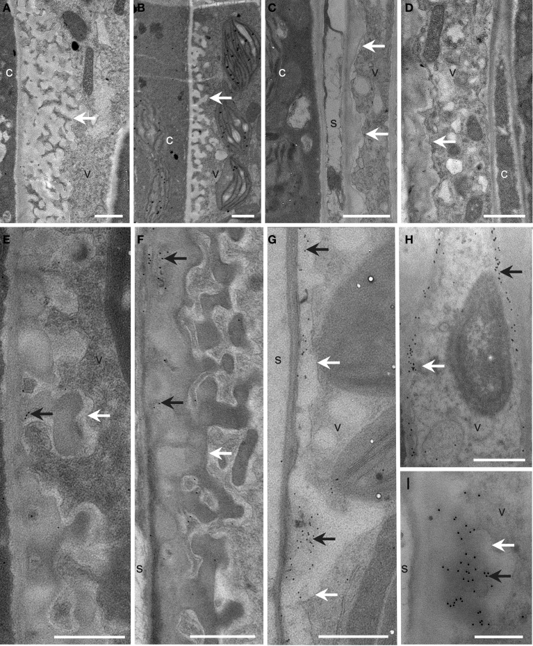
Figure 6.
Cellular structure and immunodetection of callose after 3 days of LT treatment. Col, gsl5, vte2, and gsl5 vte2 were grown under permissive conditions for 4 weeks and transferred to LT conditions for 3 additional days. Col (A,E), gsl5 (B,F), vte2 (C,G), and gsl5 vte2 (D,H,I). Black arrows highlight wall ingrowths of phloem parenchyma transfer cells immunolabeled with anti-β-1,3-glucan. White arrows mark transfer cell walls. c, companion cell; s, sieve element; v, vascular parenchyma transfer cell. Bars = 1 μm (A–H), 0.5 μm (I).

