Figure 1.
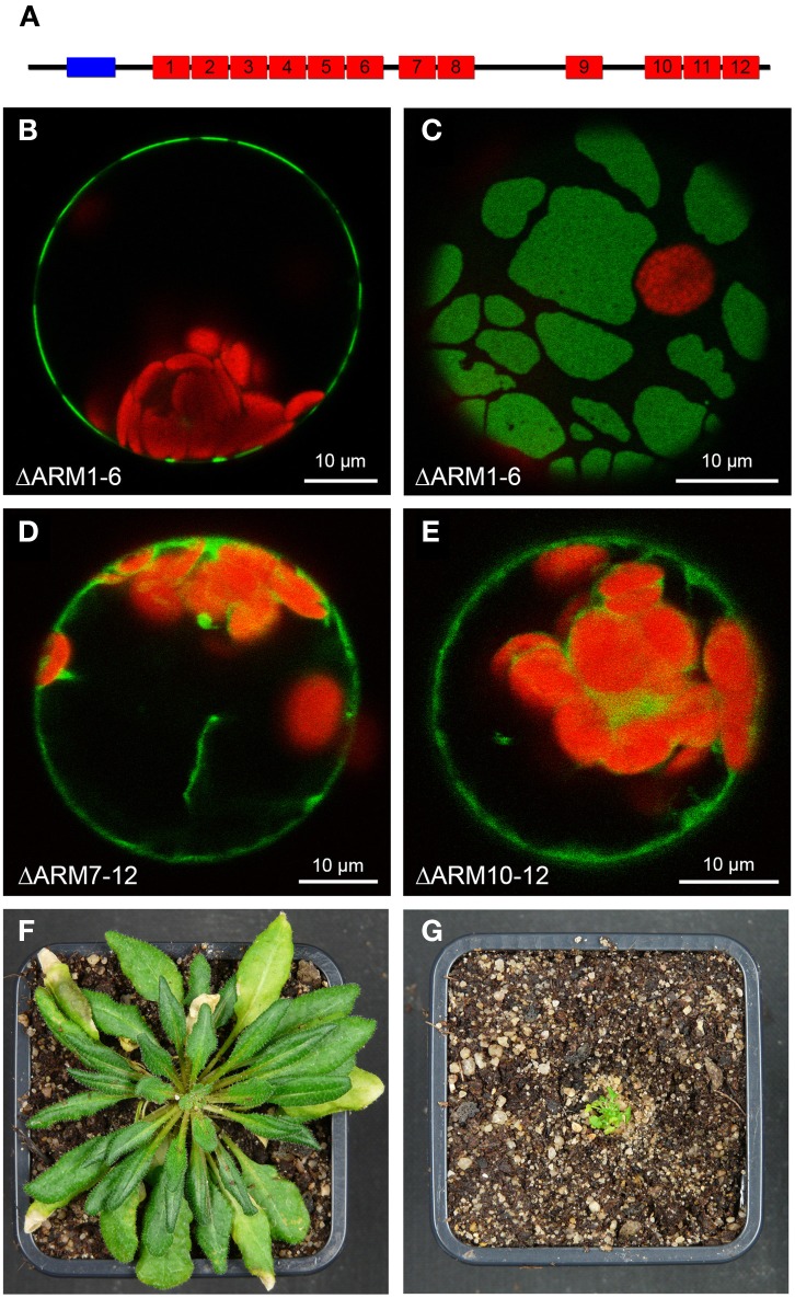
Involvement of C-terminal ARM repeats in plasma membrane-association of AtPUB43. (A) Schematic representation of the AtPUB43 protein domain structure. U-box and ARM repeats are depicted in blue and red, respectively. (B) Confocal analysis of fluorescence of an Arabidopsis leaf protoplast expressing AtPUB43ΔARM1−6-GFP fusion proteins. GFP fluorescence depicted in green was only detected at the plasma membrane. Chlorophyll auto-fluorescence within the chloroplasts is depicted in red. (C) Confocal analysis of fluorescence of an Arabidopsis leaf protoplast expressing AtPUB43ΔARM1−6-GFP fusion proteins. The representative top-view showed that GFP signals and thus the AtPUB43ΔARM1−6-GFP fusion proteins were localized in membrane patches. (D) Confocal analysis of fluorescence of an Arabidopsis leaf protoplast expressing AtPUB43ΔARM7−12-GFP fusion proteins. GFP fluorescence depicted in green was detected in the cytosol. Chlorophyll auto-fluorescence within the chloroplasts is depicted in red. (E) Confocal analysis of fluorescence of an Arabidopsis leaf protoplast expressing AtPUB43ΔARM10−12-GFP fusion proteins. GFP fluorescence depicted in green was detected in the cytosol. Chlorophyll auto-fluorescence within the chloroplasts is depicted in red. (F) Growth phenotype of wildtype plants grown for 12 weeks in short-day conditions. Photon flux density was 80–100 μmol m−2 s−1. (G) Growth phenotype of CaMV35S::YFP-SAUL1 plants grown for 12 weeks in short-day conditions. Photon flux density was 80-100 μmol m−2 s−1.

