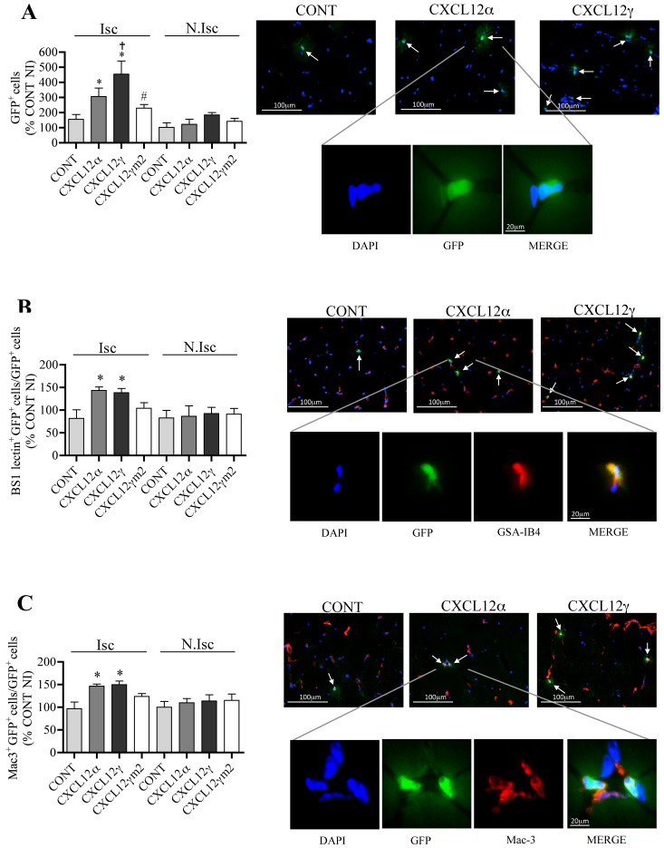
Figure 6. CXCL12γ controls BM-derived cells infiltration in ischemic tissues.
Quantitative evaluation and representative photomicrographs of the percentage of GFP-expressing cells (A), GFP-expressing and BS1-labelled cells (B) and GFP-expressing and Mac3-labelled cells (C) in the ischemic leg of irradiated wild-type mice reconstituted with bone marrow of WT GFP+ animals and treated with empty DNA plasmid (CONT), or DNA expression vectors encoding for CXCL12α, CXCL12γ or HS-binding mutant CXCL12γm2. Value quantification are represented in histograms. Values are mean ± SEM. n = 10 per group, representative of 2 independent experiments. A–C) 28 possible comparisons for Bonferroni correction, a value of p<0.0017 was considered significant, *p<0.0017 versus Isc CONT, †p<0.0017 versus Isc CXCL12α, #p<0.0017 versus Isc CXCL12γ. Isc indicates ischemic leg and N.Isc, non ischemic leg.

