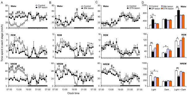
Fig. 7.
Quantitative changes of sleep–wake times of ibotenic acid lesions in globus pallidus (GP), substantia nigra pars reticulate (SNr) and subthalamic nucleus (STN). (A–C) Diurnal patterns of hourly wake, rapid eye movement (REM) and non-rapid eye movement (NREM) sleep amounts of control (n = 6) and GP (n = 6), SNr (n = 5) and STN (n = 6) lesion groups, *P < 0.05, **P < 0.01, two-tailed unpaired t-test. (D) Total amounts of wake, REM and NREM sleep in light period, dark period and light + dark period in each group. *P < 0.05, **P < 0.01, one-way ANOVA followed by Dunnett’s post hoc test.

