
Fig. 9.
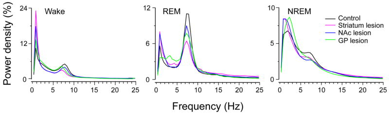
EEG power spectra during wake, rapid eye movement (REM) and non-rapid eye movement (NREM) sleep over 24 h. The power spectrum was normalized to total power (0.5–24.5 Hz). Lesions in the striatum, nucleus accumbens core (NAc) and globus pallidus (GP) all produced a generalized slowing of the EEG, with less theta and more delta density during wake, REM and NREM sleep. In other words, all of the lesions produced a slowing of the cortical EEG.