Figure 7.

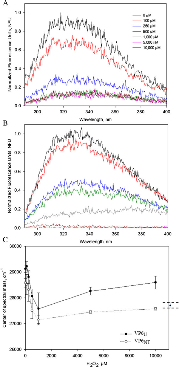
Emission fluorescence scans at λex 295 nm of VP6NT and VP6U (0.4 mg/mL) after MCO with 150 µM of FeCl2 at various H2O2 concentrations for 1 h. A) Normalized emission spectra of VP6NT. B) Normalized emission spectra of VP6U. C) The center of spectral mass (CSM) of oxidized VP6NT and VP6U was calculated using Equation (3) and plotted against the H2O2 concentration used. On the right, the CSM of denatured VP6 (treated for 2 hours with 6 M of guanidine chloride) is shown for comparison. Experiments were performed in triplicate, except for the CSM of denatured VP6, which was analyzed in duplicate. Error bars represent the standard deviation or difference among them.
