
Figure 2.
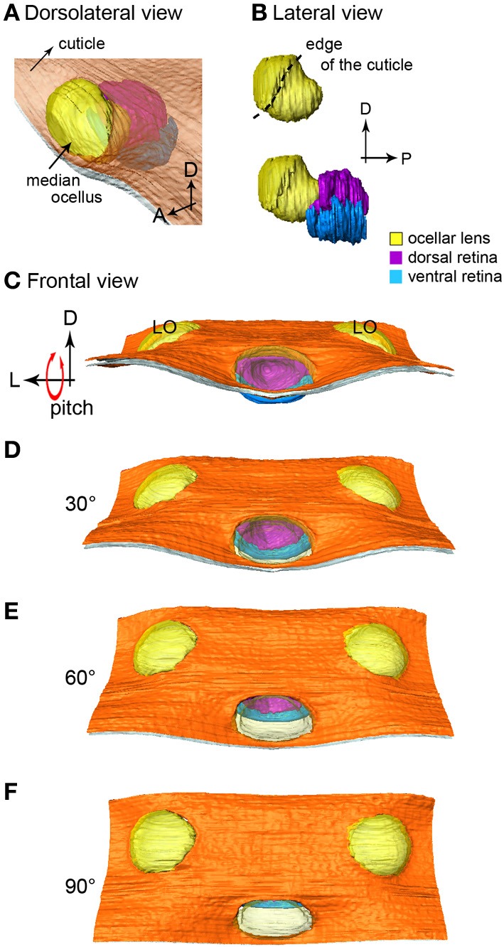
The 3-D reconstruction of the honeybee median ocellus. (A) Dorso-lateral view of the median ocellus with the cuticle in view. (B) Lateral views of the median ocellar lens (l, shown in yellow) and dorsal and ventral retinas. The lens of the median ocellus is elongated downward, the retinas are positioned downwards and posteriorly in relation to the lens. (C–F) Simulation of a pitching movement of the model. (C) A frontal view of the median ocellus: only the dorsal retina can be seen through the median ocellar lens (the ventral retina is under the cuticle). (F) A dorsal view of the ocelli: only the ventral retina can be seen. Abbreviations: D, dorsal; L, lateral; LO, lateral ocellus; P, posterior.