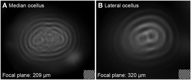
Figure 4.

Imaging of concentric circles through the median (A) and lateral ocellar (B) lenses of the honeybee. Both (A,B) show images of angularly corrected stimuli of 5° spatial wavelength on a wide-field LCD monitor as seen through the lenses at the focal plane. Both the median and lateral ocellar lenses form a single image but the centers of the images degrade in both cases.
