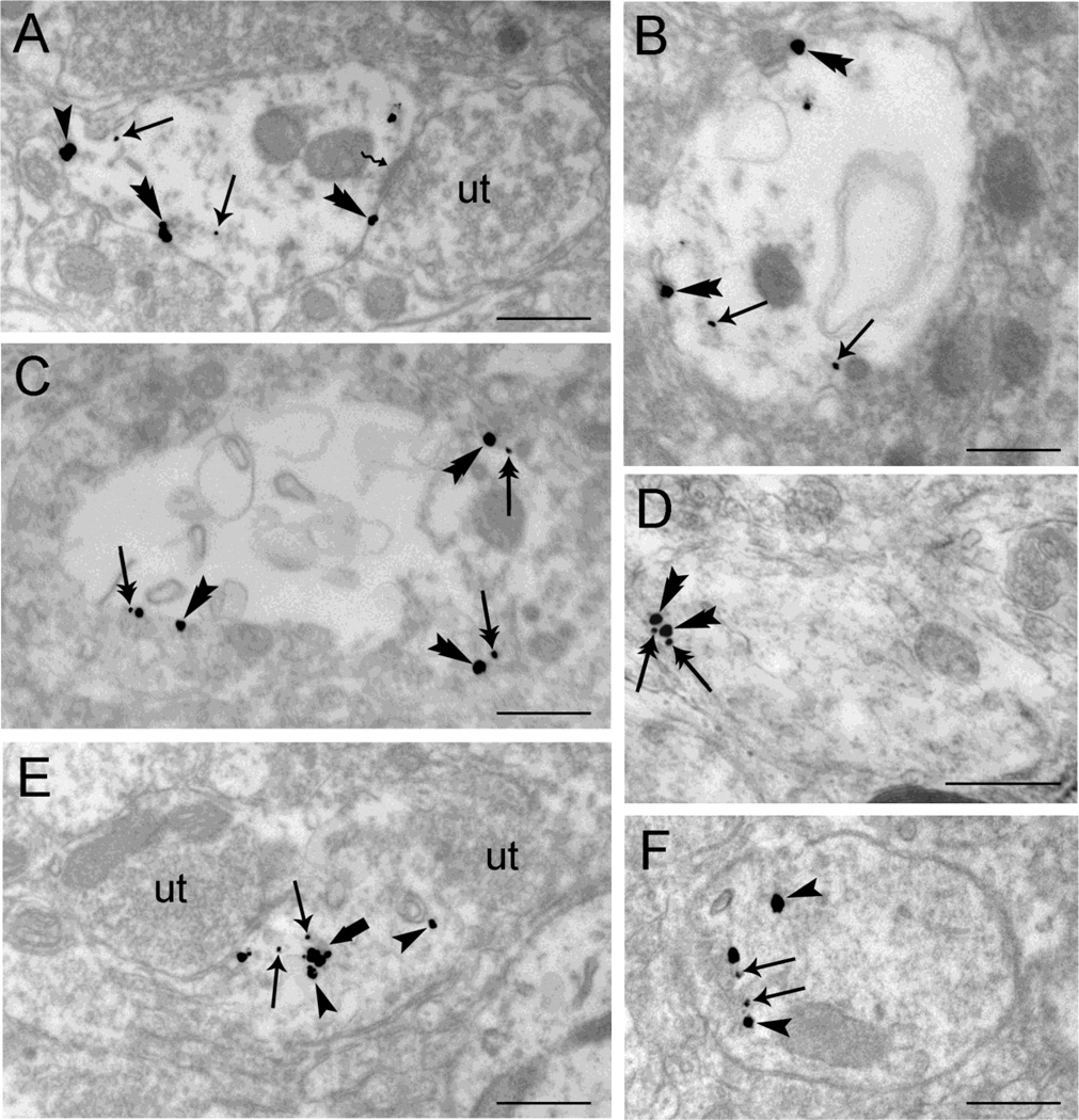
Figure 5.
Differential trafficking of WLS is observed in rat locus coeruleus (LC) dendrites from vehicle-treated (A, B), morphine-treated (C–D) and DAMGO-treated rats (E–F) using dual immunogold-silver for WLS (small gold-silver grains) and MOR (large gold-silver grains). A-B. Immunogold-silver labeling for WLS can be seen within the cytoplasm (arrows) while immunogold-silver labeling for MOR is distributed within the cytoplasm (arrowhead) and along the plasma membrane (double arrowheads) in dendrites from vehicle-treated rats. Panel A shows an unlabeled axon terminal (ut) contacting (zigzag arrow) the WLS and MOR-containing dendrite. C-D. Following morphine treatment, WLS labeling shifts from the cytoplasm to the plasma membranes of LC dendrites. Double arrows point to immunogold-silver labeling for WLS along the plasma membranes while double arrowheads point to immunogold-silver for MOR labeling along the plasma membrane in WLS and MOR-containing dendrite. E-F. Immunogold-silver labeling for WLS (arrows) and MOR (arrowheads) can be seen primarily within the cytoplasm of the WLS and MOR-containing dendrites in rats subjected to DAMGO treatment. Two unlabeled axon terminals (ut) can be seen contacting a WLS and MOR-containing dendrite (Panel E). A thick arrow points to endosome-like vesicles. Scale bars, 0.5µm.

