Figure 1.
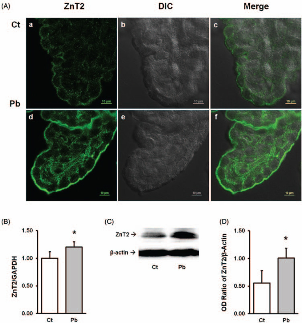
Expression of ZnT2 in choroid plexus tissue and effect of Pb exposure. A(a–c). Choroid plexus tissues were dissected from the lateral ventricles. A(d–f). For in vivo Pb exposure, animals received a single ip injection of 50 mg PbAc for 24 h. A(a) and A(d) show the ZnT2 green fluorescent signal; A(b) and A(c) show the DIC mages; A(c) and A(f) are the merged images of ZnT2 fluorescent signal and DIC. (B) qPCR quantitation of ZnT2 mRNA expression in the choroid plexus. Data represent mean ± SD, n=6, *P < 0.05, as compared with the control. (C) Western blot analysis of ZnT2 in the choroid plexus. β-actin was used as the house-keeping protein. (D) Quantification of western blot was expressed as the OD ratio of ZnT2/β-actin. Data represent mean ± SD, n=3, *P < 0.05 as compared with the control. (A color version of this figure is available in the online journal.)

