
Figure 1.
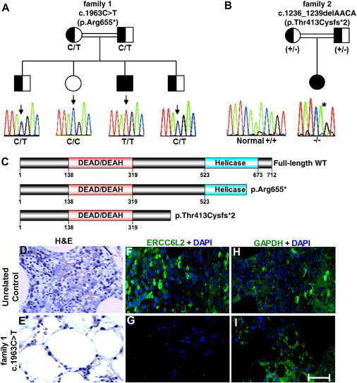
Truncating Mutations in ERCC6L2
(A and B) Shown are two families in which ERCC6L2 mutations segregate as an autosomal-recessive trait. A Sanger sequencing trace and the genotype of each individual are given; inferred genotypes are in parentheses.
(B) For family 2, a plus sign (+) indicates the WT allele and a minus sign (−) indicates the mutant allele. The normal sequencing trace to the left of family 2 comes from an unrelated individual.
(C) The identified ERCC6L2 alterations leading to premature truncation are indicated on a diagram of the protein; functional domains are also annotated.
(D and E) H&E staining of bone marrow trephine biopsies from an unrelated control sample and the index case (case 1) from family 1 reveals the degree of hypoplasia in this individual.
(F and G) Immunostaining on these bone marrow trephine sections revealed the presence of ERCC6L2 in a normal unrelated control, but it was not detected in the affected individual.
(H and I) Positive control staining for GAPDH antigen was observed in both sections. Figures are representative of different images taken from different fields of view. Magnification is 40×. The scale bar represents 50 μm.