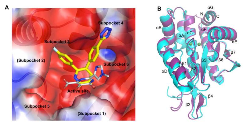
Figure 1.
Description of PAN structure. (A) APBS51 calculated electrostatic potential surface of the active site cleft of PAN. The binding of a ligand described in this study (7) is shown with yellow carbon atoms and a carboxamide derivative with three chelating groups crystallized by Dubois et al. is shown in gray (PDB code: 4E5I).19 The subpockets are labeled as previously described.19 (B) Superposition of the pH1N1 2009 PAN structures from this study and one previously described (PDB code: 4AVQ).18

