
FIG. 2.
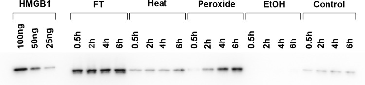
The kinetics of HMGB1 release by necrotic cells. In these experiments, Jurkat T cells were cultured in serum-free medium and induced to undergo necrosis by the following treatments: freeze–thaw, heat, hydrogen peroxide (H2O2), ethanol, or no treatment (controls). After treatment, cells were incubated at 37°C for the times indicated. Culture media were concentrated and then examined by western blotting using a mouse monoclonal anti-HMGB1 antibody followed by a horseradish peroxidase-conjugated goat anti-mouse reagent and detection by enhanced chemiluminescence. Controls for HMGB1 detection (100, 50, and 25 ng of recombinant hHMGB1-histidine tagged protein) were also analyzed. Figure is a digital image of a representative blot. As these data indicate, the release of HMGB1 differs significantly depending on the treatment to cause necrosis. Reproduced with permission from Beyer et al. (8).