
Fig. 2.
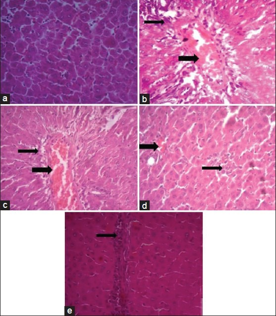
Effect of D. salina on the liver histopathological photomicrographs of the experimental groups of rats.
Histopathological photomicrographs (×400) of livers of various groups stained with haematoxylin and eosin. (a) Normal architecture of rat liver, (b) Necrosis and hepatocellular fatty degeneration (eccentric nuclei) in acetaminophen intoxicated liver and congestion of portal vein and peri-portal infiltration of inflammatory cells, (c) Lesser damage of hepatocytes and low index of necrosis (centrally located nuclei) in D. salina-500 mg/kg pretreated group, (d) Minimal damage of hepatocytes and very low index of necrosis in D. salina-1000 mg/kg pretreated group and mild congestion, (e) Very lesser damage of hepatocytes and low index of necrosis in silymarin pretreated group, narrow arrows refer to inflammatory cells infiltration, wide arrows refer to congestion of portal vein.