
Figure 3.
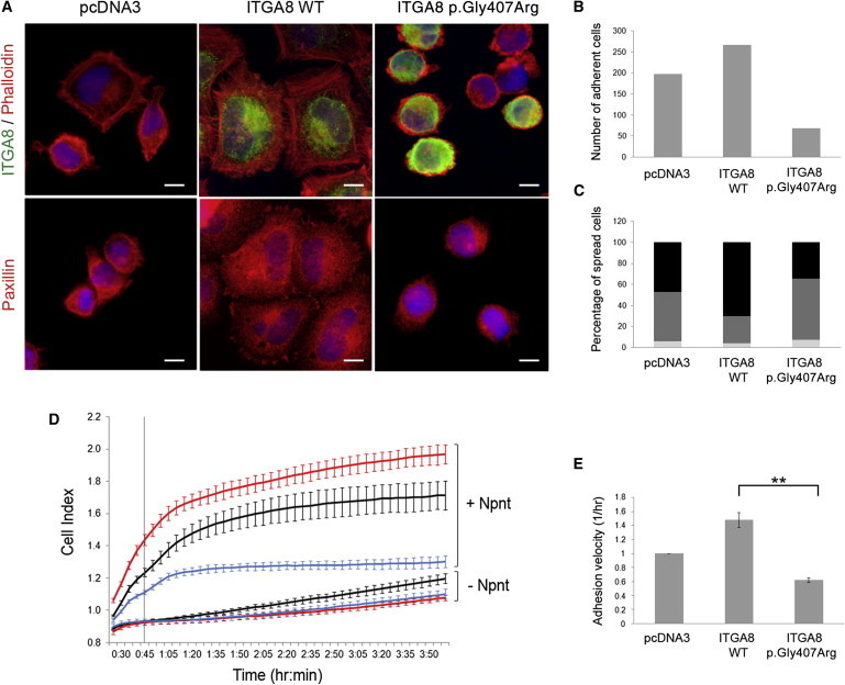
Characterization of the Effect of the ITGA8 p.Gly407Arg Variation on Cell Adhesion and Spreading
(A–C) Analysis of adhesion and spreading by immunofluorescence.
(A) For each cell line, 2 × 10⁵ cells were plated on glass slides previously coated with nephronectin. After a 45 min incubation at 37°C, cells were fixed with PFA 4%. Staining of ITGA8 (rabbit anti-integrin α8 1/100), filamentous actin (rhodamine coupled phalloidin 1/400, Sigma-Aldrich), and paxillin (mouse anti-paxillin 1/50, BD Biosciences) showed enhanced spreading of ITGA8 WT cells compared to ITGA8 p.Gly407Arg cells.
(B) Cell adhesion was measured by counting the number of adhered cells in 12 fields (at magnification ×63) for each cell line.
(C) The percentage of spread and unspread cells was estimated by counting of 100 cells for each cell line (light gray, unspread; medium gray, weakly spread; black, strongly spread).
(D and E) Lifetime cell adhesion and spreading was evaluated with the impedance-based system xCELLigence (ACEA Biosciences, Roche). E-type plates were coated for 1 hr at 37°C with 10 μg/ml of nephronectin (Npnt) and rinsed for an additional 1 hr with 1% BSA/PBS. 8 × 10⁴ stably transfected HEK293 cells (ITGA8 WT, ITGA8 p.Gly407Arg, or empty pcDNA3) were seeded in each well.
(D) Cell index (CI) was measured from 25 min after seeding. Each CI value corresponds to the average of triplicates ± standard deviation (red curves, ITGA8 WT; blue curves, ITGA8 p.Gly407Arg; black curves, empty pcDNA3).
(E) Adhesion velocity is represented as the slope of the curves, calculated on the first 45 min after seeding (indicated by the vertical line in D). The graph represents the mean ± SEM of two independent experiments, each one in triplicate. ∗∗p < 0.01 calculated by t test.