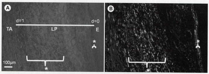
Fig 2.

A) Coronal section of human vocal fold observed under circularly polarized light. TA — thyroarytenoid muscle; LP — lamina propria; E — epithelial layer; d — normalized depth of LP; d=0 — E/LP boundary; d=1 — TA/LP boundary. B) Birefringence values of image in A obtained from Metripol system, where white indicates strong birefringence. Bracketed areas and asterisks indicate regions with high birefringence within LP.
