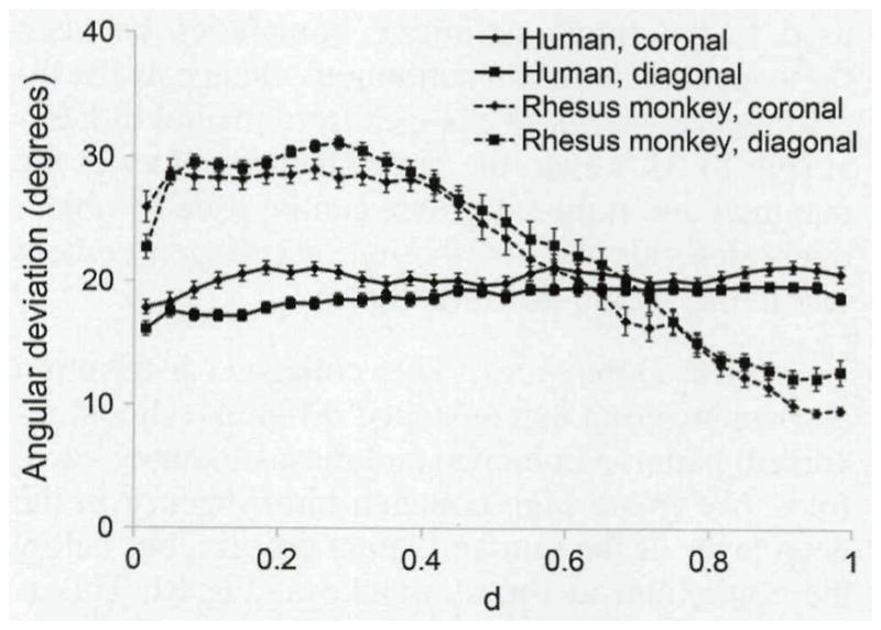
Fig 7.

Mean (±SD) angular deviations at various depths within lamina propria. Fiber organization was more consistent throughout thickness of human samples. In rhesus monkey samples, fibers were much more disorganized closer to epithelial layer. Angular deviation of zero indicates perfectly aligned fibers. At 40.5°, fibers are completely randomized. TA — thyroarytenoid muscle; LP — lamina propria; E — epithelial layer; d — normalized depth of LP; d=0 — E/LP boundary; d=l — TA/LP boundary.
