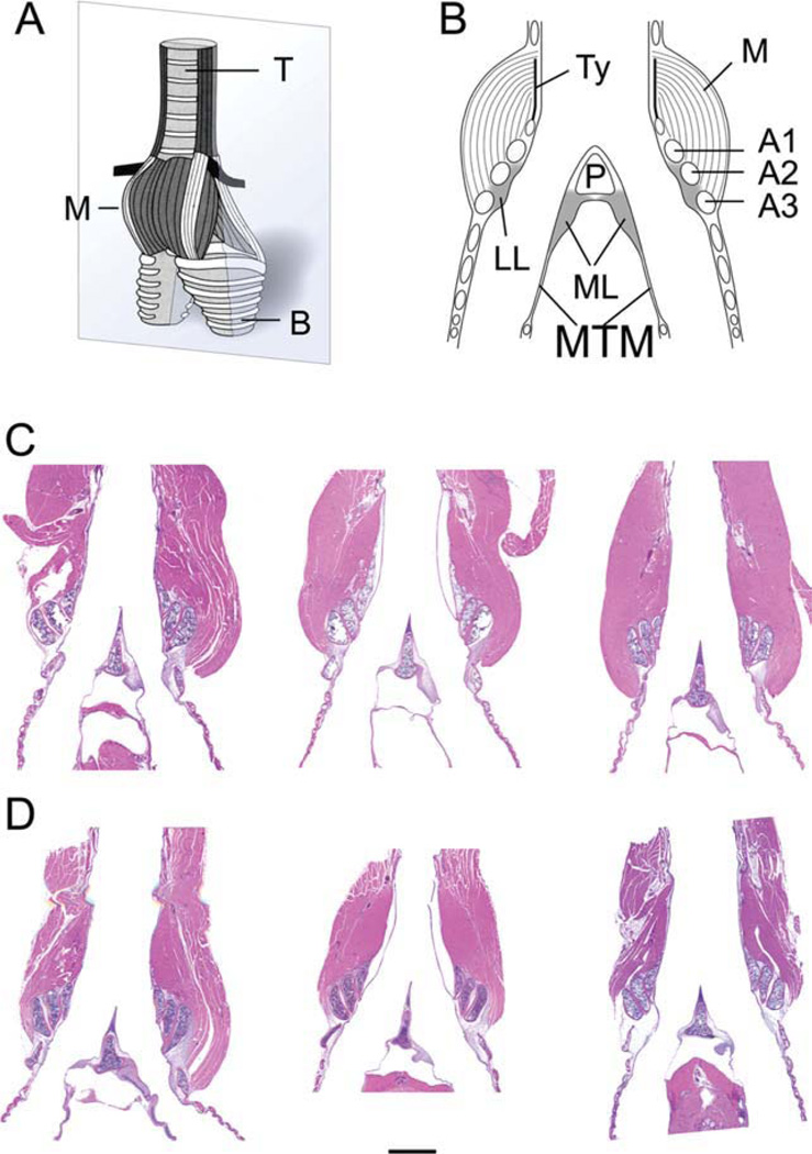
Fig. 1.
A: Schematic ventral view of a syrinx. The grey plane indicates the section level of the other images (B–D). B: Schematic of a mid-organ section through syrinx indicating all elements measured. C: Mid-organ section of three male syringes (HandE stain). D: Mid-organ section of three female syringes (HandE stain). Note the smaller muscle mass in females. The bar in D indicates a 1 mm distance and applies to C and D. M, intrinsic syringeal muscles; B, primary bronchi; T, trachea; LL, lateral labia; ML, medial labia; MTM, medial tympaniform membrane; Ty, tympanum; P, pessulus; A1, A2, A3, first, second and third bronchial half ring.

