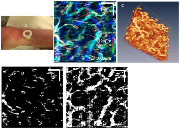
Fig. 2.
A: Photograph shows the imaging location (area enclosed by white circle) on a subject’s PWS. B: The MIP images of the PWS skin microvasculature. C: 3D rendering of the PWS skin microvasculature (movie). D: PWS skin microvasculature at a layer close to the epidermal–dermal junction. E: PWS skin microvasculature at a depth approximately 800 μm below the skin surface.

