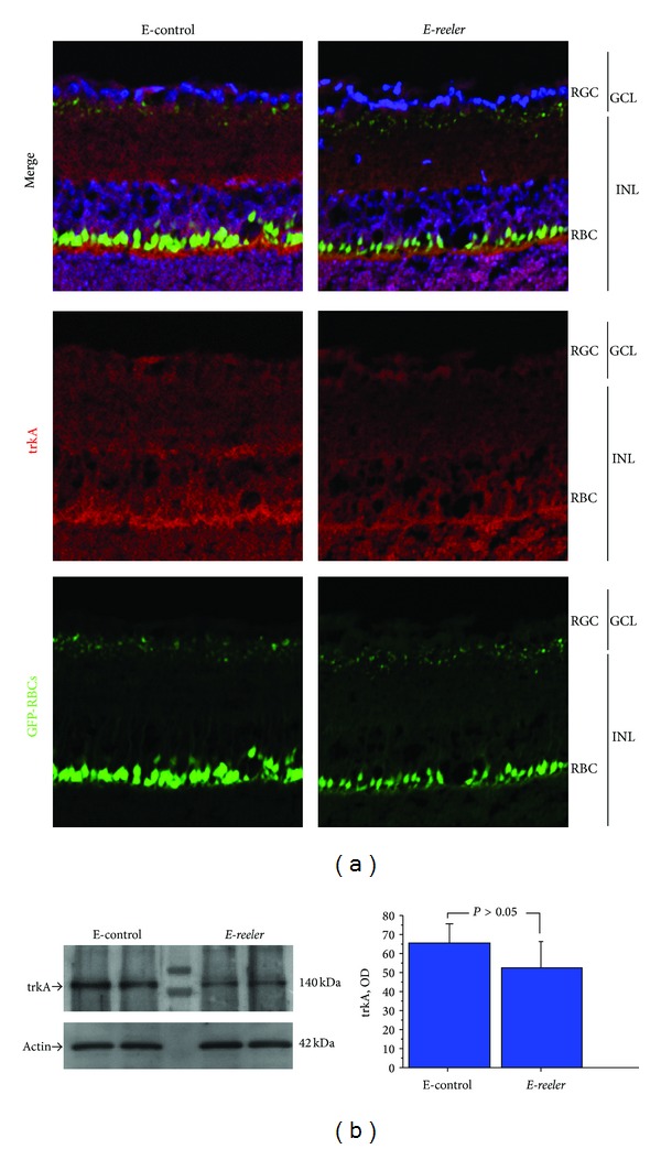
Figure 3.

Expression of trkANGFR in E-reeler retina. (a) Confocal microscopy showing images of GFP-expressing RBCs (green), trkANGFR immunoreactivity (red), and nuclear staining (blue). A weak trkANGFR immunoreactivity was observed at the RBCs (body and dendrites). trkANGFR staining was less intense across the INL and in some cells inside the GCL of the E-reeler retina (×400). (b) Representative 7.5% SDS-PAGE and relative densitometric analysis of E-control and E-reeler retinal extracts probed with the trkANGFR antibody (OD values; P > 0.05). The size-marker was run between the two groups. Abbreviations: GFP, Green Fluorescent Protein; RGC, Retinal Ganglion Cells; GCL, Ganglion Cell Layer; INL, Inner Nuclear Layer; RBCs, Rod Bipolar Cells; OD, Optical Density.
