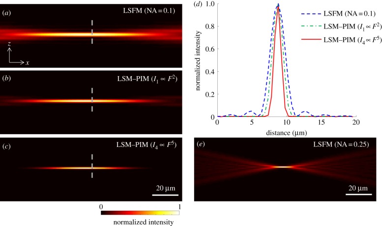
Figure 2.
Simulated effective excitation volume in (a) LSFM with an excitation objective of NA = 0.1, (b) the corresponding first-order LSF–PIM and (c) fourth-order LSF–PIM. (d) Comparison of signal intensities along the dashed line in (a–c). (e) Simulated effective excitation volume in LSFM with an excitation objective of NA = 0.25. The optical sectioning thickness in (c) is comparable to that in (e), yet with a six times larger (in diameter) imaging FOV. (Online version in colour.)

