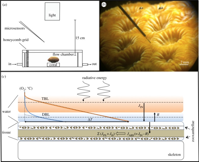
Figure 1.
Experimental setup. (a) Schematic of the experimental setup visualizing the relative position of light source, microsensors and coral fragment. (b) A scalar irradiance microsensor inserted into the coenosarc tissue of a M. curta coral. (c) Conceptual diagram showing the fate of light energy (abbreviations explained in text).

