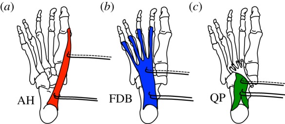
Figure 2.

Location of electrodes within the intrinsic foot muscles. Schematic of the anatomical location of (a) abductor hallucis (AH), (b) flexor digitorum brevis (FDB) and (c) quadratus plantae (QP) from the plantar aspect of a right foot. Fine wire pairs of electromyography (EMG) electrodes (black lines with hooked ends) were inserted under ultrasound guidance, with one pair being inserted proximally and one pair distally to the muscle belly. The proximal electrode pair was used for the EMG recordings in experiment 1, whereas one wire from each of the proximal and distal pairs was connected to a constant current electrical stimulator, which delivered trains of electrical stimulation to each muscle independently in experiment 2. (Online version in colour.)
