Figure 1.
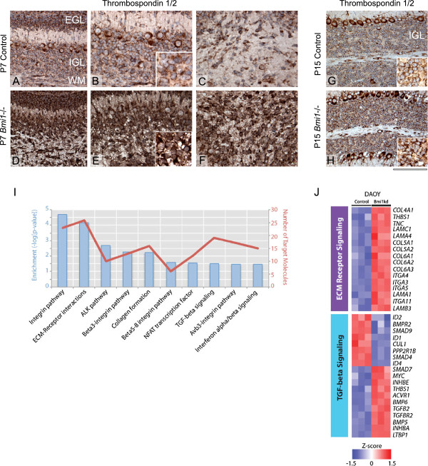
BMI1 regulates the expression of cell adhesion genes in GCPs and ECM remodelling and TGFβ signalling in DAOY MB cells. (A-H) Thrombospondin 1/2 immunohistochemistry on murine P7 cerebella show increased expression in GCPs in Bmi1-/- mice (D and E) compared to the control cerebellum (A and B), higher magnification is shown in the inset. Increased expression of Thrombospondin 1/2 is also seen in the white matter glial cells of Bmi1-/- (F) compared to P7 controls (C). Granule cells in the IGL of Bmi1-/- P15 mice also display increased Thrombospondin 1/2 expression (H) as compared to control (G), inset show higher magnification. (I) Molecular Signature Database (MSigDB) analysis of genes differentially expressed between DAOYBMI1kd versus control cells identifies significantly (FDR q < 0.05) over-represented pathways [25]. (J) Heat map representation of the relative upregulation of ECM receptor signalling genes (upper panel) and silencing of TGF-beta signalling (lower panel) in DAOYBMI1kd compared to control cells. Genes were identified using MSigDB and plotted to highlight differences in expression of 3 z-scores or greater between the two groups (red = relative upregulation, blue = relative downregulation). EGL: External granular layer, IGL: Internal granular layer, WM: White matter. Scale bar is 125 μm (A, D, G, H) and 80 μm (B, E, C, F) and 50 μm (B inset, E inset, G inset, H inset).

