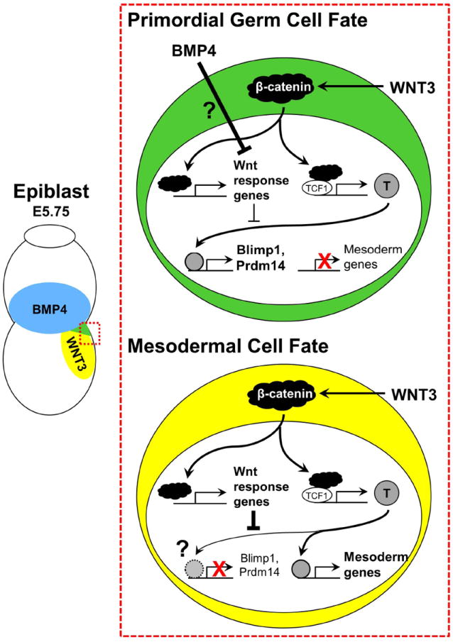
Figure 1. Model of PGC/mesoderm fate choice.

Mouse PGCs are specified in the posterior corner of the proximal epiblast where Bmp4 (blue) and Wnt3 (yellow) signals converge. Neighboring mesodermal cells do not receive high levels of Bmp signals, preventing their specification to the germ cell lineage.
