Figure 3.
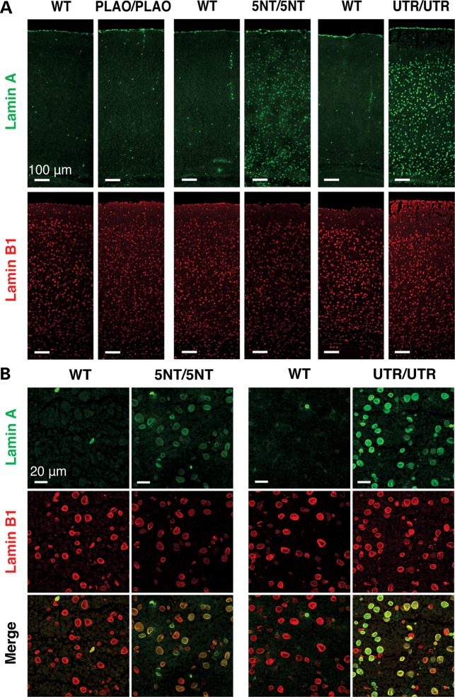
Immunofluorescence microscopy of the cerebral cortex from Lmnaplao/plao (PLAO/PLAO), Lmnaplao-5nt/plao-5nt (5NT/5NT), Lmnaplao-utr/plao-utr (UTR/UTR) and wild-type (WT) littermate mice, revealing increased lamin A expression in the mice harboring mutations in prelamin A's 3′ UTR. (A and B) Low- and high-magnification images of the cerebral cortex stained with antibodies against lamin A (green) and lamin B1 (red). The scattered lamin A-positive cells in wild-type and Lmnaplao/plao mice are endothelial cells (5). Scale bar in (A), 100 µm; (B), 20 µm.

