Figure 4.
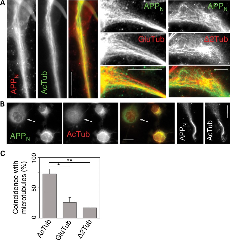
The NTFs of APP primarily associate with acetylated microtubules in CAD cells. (A) Significant co-localization of APP N-terminal epitopes (APPN; detected with antibody 22C11) with acetylated microtubules (AcTub; detected with monoclonal antibody 611B1), but not with detyrosinated (GluTub) or delta 2-tubulin (Δ2Tub) microtubules. (B) Extensive co-localization of APPN with acetylated microtubules (detected with polyclonal anti-acetyl-K40 antibody) in the soma, as well as at the neurite terminals. Note that the perinuclear accumulation of NTFs is largely diminished in the cell lacking a perinuclear network of acetylated microtubules (arrow). Scale bars, 20 µm. (C) Quantitative analysis of co-localization of APPN with microtubules showing different posttranslational modification. In spite of a high cell-to-cell variability, the coincidence of the 22C11 immunolabeled filaments with AcTub is significantly higher than that with GluTub (* indicates P < 0.01) or Δ2Tub (** indicates P < 0.001). Bars represent SEM.

