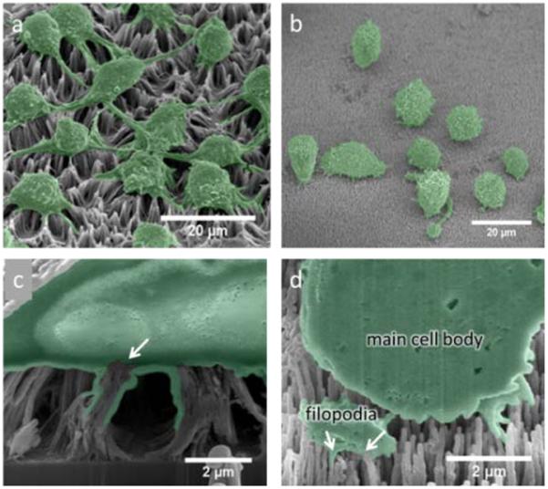
Fig. 3.

(a) Representative SEM image of DC 2.4 cells on a bare CNT array and (b) on an amorphous alumina-coated CNT array. (c) FIB cross-section of a DC 2.4 cell cultured on a CNT array, the arrow indicates point of CNT engulfing resulting in a broken membrane, (d) FIB cross-section of a DC 2.4 cell on an alumina-coated CNT array, arrows indicate engulfing of individual alumina NWs through a sectioned portion of filopodia.
