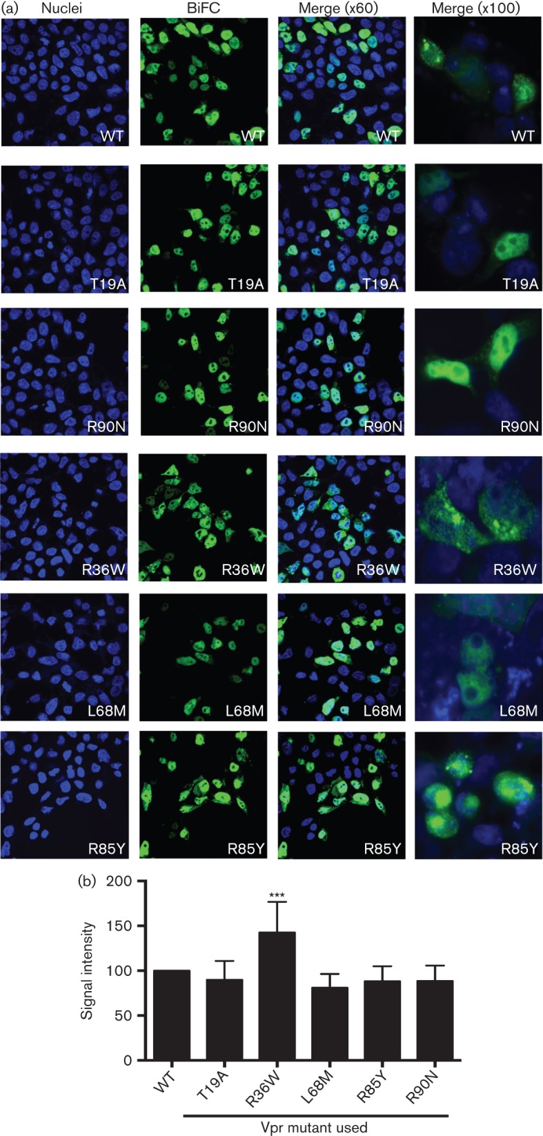
Fig. 4.

Subcellular localization of Vpr variants by microscopy. (a) The subcellular localization of the Vpr variant oligomers was assessed by co-transfection of the VC-Vpr and VN-Vpr mutant constructs in HEK293T cells. Thirty-six hours post-transfection cells were fixed, stained with DAPI for visualization of nuclei, and visualized under fluorescence microscopy at magnifications of ×60 and ×100 to assess the localization of the BiFC signal within the cell. The figure represents one of six independent experiments. Blue, DAPI, represents nuclei; green, BiFC, represents Vpr oligomers. (b) BiFC signal was quantified using NIS Elements Ar Microscope Imaging Software. BiFC positive cells (n = 12) per field were quantified from multiple fields and intensity was measured in total cells, cytoplasmic and nuclear compartments. Signal in wild-type (VprNL43) was normalized as 100 % and the mutants were calculated accordingly.
