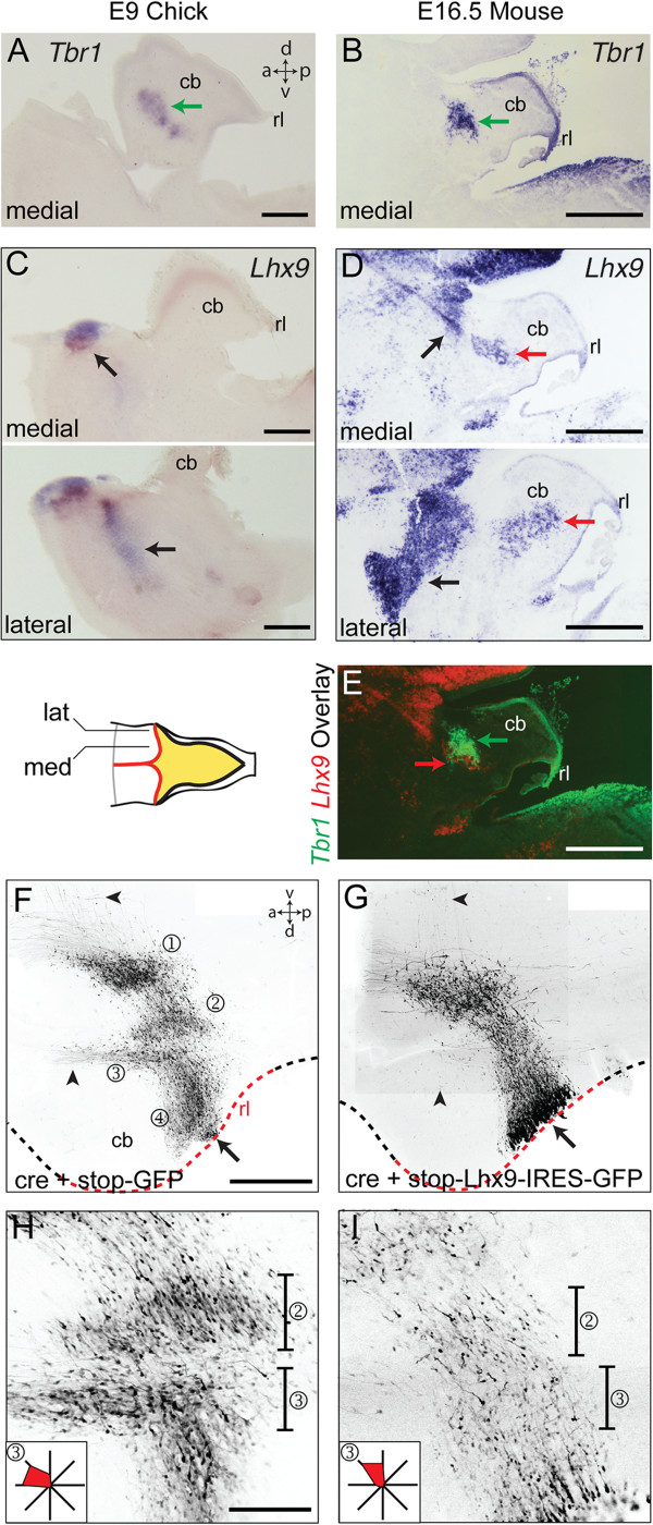
Figure 4.

Lhx9 overexpression in chicks alters nucleogenesis and initial axon trajectory of rhombic lip derivatives. A, B. Medial/fastigial nucleus expression of Tbr1 in sagittal sections of st.35/E9 chicks (A) and E16.5 mice (B). C. Lhx9 expression in medial (above) and lateral (below) sagittal section of st.35/E9 chicks in extra-cerebellar populations (black arrows). Since these are lateral sections, the area of cerebellar tissue is smaller than mid-sagittal sections. D. Lhx9 expression in medial (above) and lateral (below) sagittal section of E16.5 mouse in both extra-cerebellar populations (black arrows) and dentate nucleus (red arrows). E. Overlay of B and D showing close apposition of Tbr1 (green arrow) and Lhx9 (red arrow) labelled neurons in cerebellar nuclei. F, G. Flat-mounted embryos at st.31/E7 electroporated at st.24/E4 with either cre + stop-GFP (F) or cre + stop-Lhx9-IRES-GFP (G). Arrowheads indicate the position of axons from cerebellar nuclei (F), which are absent following Lhx9 overexpression (G). Single optical sections shown respectively in H and I show lateral (②) and medial (③) cerebellar nuclei at high magnification with vectors of initial cell processes (in region ③) shown inset as relative frequencies on a radial plot. Scale bars: 500 μm in A-E and F-G, 200 μm H-I.
