
Figure 3. PanIN-like lesions developed within the first two weeks after birth when sHB-EGF was overexpressed in conjunction with Kras activation.
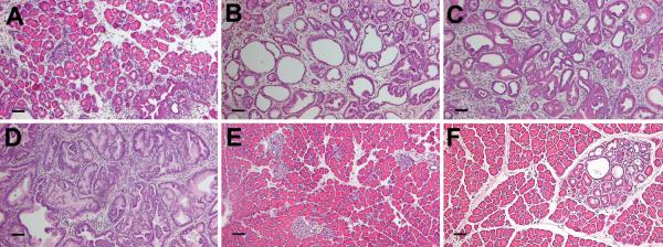
KrasG12D; sHB-EGF mice (A–D), KrasG12D alone (E) and sHB-EGF alone (F) were analyzed at 1 day (A), 3 days (B), 7 days (C) and 14 days (D–F) after birth by hematoxylin (blue) and eosin (pink) staining. A. In KrasG12D; sHB-EGF mice on postnatal day 1, some acinar and ductal lumena were slightly dilated. By postnatal day 3, large cystic ducts with columnar cell morphology were apparent with few acini remaining. By day 7, no structural acini remained and cells within ducts became columnar with occasional papillary architecture and lumenal budding observed. By 14 days, these structural alterations and other cellular changes were consistent throughout each KrasG12D; sHB-EGF pancreas while no or few alterations were observed with KrasG12D or HB-EGF alone. Size bars, 50 μm.