
Figure 4. Overexpression of sHB-EGF in conjunction with activated Kras resulted in acinar-ductal metaplasia of most exocrine cells.
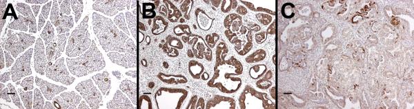
A. Normal pancreas at postnatal day 14 was comprised largely of acinar cells with a smaller number of duct cells as visualized by CK19 immunolabelling (brown). B. By day 14 in KrasG12D; sHB-EGF mice, CK19 labeled the majority of epithelial cells in the pancreas. C. Immunolabeling for amylase (brown) revealed that no acinar structures remained in KrasG12D; sHB-EGF mice and only rare amylase-positive acinar cells were found within ductal structures. Many fields of view completely lacked amylase-positive cells (data not shown). Size bars, 50 μm.